Effect of the Innominate Bone Horizontal Rotation on Acetabular Version: A Retrospective Radiological Study on a Middle Eastern Population
DOI:
https://doi.org/10.38179/ijcr.v3i1.164Keywords:
Acetabular version, Computed tomography, Hemipelvis, Hip joint, Horizontal rotation, Innominate boneAbstract
Background: The impact of acetabular horizontal rotation on the development of femoroacetabular impingement and subsequently osteoarthritis is well-studied in the literature. However, there is not a clear relationship between the rotation of the hemipelvis and the version of the acetabulum.
Purpose: The purpose of this study was to evaluate the influence of the rotation of the hemipelvis on the version of the acetabulum.
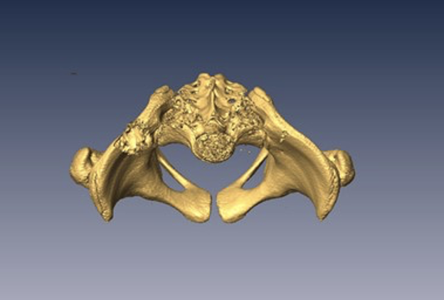
Methods: Through a retrospective study, three-dimensional reconstructions of high-resolution CT (computed tomography) scans of 154 patients receiving pelvic scans for non-orthopedic causes were selected from our institution’s database. The horizontal rotation of the different parts of the hemipelvis was evaluated using the following parameters: superior iliac spine angle (SIS), inferior iliac spine angle (IIS), roof edge angle (REA), equatorial edge angle (EEA) and ischiopubic angle (IPA).
Results: The results showed a significant positive correlation between the different angles of the innominate bone and the version of the acetabulum such as when the proximal innominate bone rotates, the cranial part of the acetabulum rotates in the opposite direction. Increased anteversion angles in females compared to males were also observed.
Conclusion: The observations suggest that, in an asymptomatic population, the acetabulum should not be considered a separate entity independent from the rest of the innominate bone and that the version of the acetabulum correlates with the rotation of the hemipelvis.
References
Krebs V, Incavo SJ, Shields WH. The anatomy of the acetabulum: what is normal? Clin Orthop Relat Res. 2009 Apr;467(4):868-75. doi: 10.1007/s11999-008-0317-1. Epub 2008 Jul 22. PMID: 18648904. https://doi.org/10.1007%2Fs11999-008-0317-1
Brinckmann P, Hoefert H, Jongen HT. Sex differences in the skeletal geometry of the human pelvis and hip joint. J Biomech. 1981;14(6):427-430. PMID: 7263735. https://doi.org/10.1016/0021-9290(81)90060-9
Govsa F, Ozer MA, Ozgur Z. Morphologic features of the acetabulum. Arch Orthop Trauma Surg. 2005;125(7):453-461. PMID: 16096799. https://doi.org/10.1007/s00402-005-0020-6
Maruyama M, Feinberg JR, Capello WN, D’Antonio JA. The Frank Stinchfield Award: Morphologic features of the acetabulum and femur: anteversion angle and implant positioning. Clin Orthop Relat Res. 2001;(393):52-65. PMID: 11764371
Lubovsky O, Peleg E, Joskowicz L, Liebergall M, Khoury A. Acetabular orientation variability and symmetry based on CT scans of adults. Int J Comput Assist Radiol Surg. 2010;5(5):449-454. PMID: 20680495. https://doi.org/10.1007/s11548-010-0521-9
Lubovsky O, Wright D, Hardisty M, Kiss A, Kreder H, Whyne C. Acetabular orientation: anatomical and functional measurement. Int J Comput Assist Radiol Surg. 2012;7(2):233-240. PMID: 21822915. https://doi.org/10.1007/s11548-011-0648-3
Beck M, Kalhor M, Leunig M, Ganz R. Hip morphology influences the pattern of damage to the acetabular cartilage. J Bone Joint Surg Br. 2005;87-B(7):1012-1018. PMID: 15972923. https://doi.org/10.1302/0301-620x.87b7.15203
Siebenrock KA, Kalbermatten DF, Ganz R. Effect of Pelvic Tilt on Acetabular Retroversion: A Study of Pelves From Cadavers. Clin Orthop Relat Res. 2003;407:241-248. PMID: 12567152. https://doi.org/10.1097/00003086-200302000-00033
Ganz R, Parvizi J, Beck M, Leunig M, Nötzli H, Siebenrock KA. Femoroacetabular impingement: a cause for osteoarthritis of the hip. Clin Orthop Relat Res. 2003;(417):112-120. PMID: 14646708. https://doi.org/10.1097/01.blo.0000096804.78689.c2
Fujii M, Nakashima Y, Sato T, Akiyama M, Iwamoto Y. Pelvic Deformity Influences Acetabular Version and Coverage in Hip Dysplasia. Clin Orthop Relat Res. 2011;469(6):1735-1742. PMID: 21203874. https://doi.org/10.1007/s11999-010-1746-1
Kumeta H, Funayama K, Miyagi S, Kita J, Hosogoe Y, Murakami J, Tokita S. (1986) Inward wing ilium of adult hip dysplasia, a characteristic cross-sectional pelvic anatomy visualized by CT [in Japanese]. Rinsho Seikeigeka, (21):67–75. PMID: 21203874. https://doi.org/10.1007%2Fs11999-010-1746-1
Suzuki S. Deformity of the Pelvis in Developmental Dysplasia of the Hip: Three-Dimensional Evaluation by Means of Magnetic Resonance Image. J Pediatr Orthop. 1995;15(6):812-816. PMID: 8543613. https://doi.org/10.1097/01241398-199511000-00016
Kojima S, Kobayashi S, Saito N, Nawata M, Horiuchi H, Takaoka K. Morphological characteristics of the bony birth canal in patients with developmental dysplasia of the hip (DDH): investigation by three-dimensional CT. J Orthop Sci. 2001;6(3):217-222. PMID: 11484113. https://doi.org/10.1007/s007760100037
Musielak B, Jóźwiak M, Rychlik M, Chen BPJ, Idzior M, Grzegorzewski A. Does hemipelvis structure and position influence acetabulum orientation? BMC Musculoskelet Disord. 2016;17(1):131. PMID: 26984181. https://doi.org/10.1186/s12891-016-0982-2
Brunner R, Picard C, Robb J. Morphology of the Acetabulum in Hip Dislocations Caused by Cerebral Palsy. J Pediatr Orthop B. 1997;6(3):207-211. PMID: 9260651. https://doi.org/10.1097/01202412-199707000-00010
Kim HT, Wenger DR. Location of Acetabular Deficiency and Associated Hip Dislocation in Neuromuscular Hip Dysplasia: Three-Dimensional Computed Tomographic Analysis. J Pediatr Orthop. 1997;17(2):143-151. PMID: 9075086. https://doi.org/10.1097/00004694-199703000-00002
Werner CML, Copeland CE, Ruckstuhl T, et al. Radiographic markers of acetabular retroversion: correlation of the cross-over sign, ischial spine sign and posterior wall sign. Acta Orthop Belg. 2010;76(2):166-173. PMID: 20503941
Suzuki D, Nagoya S, Takashima H, Tateda K, Yamashita T. Three-dimensional orientation of the acetabulum. Clin Anat. 2017;30(6):753-760. PMID: 28631289. https://doi.org/10.1002/ca.22945
Giori NJ, Trousdale RT. Acetabular Retroversion is Associated With Osteoarthritis of the Hip. Clin Orthop Relat Res. 2003;417:263-269. PMID: 14646725. https://doi.org/10.1097/01.blo.0000093014.90435.64
Jamali AA, Mladenov K, Meyer DC, et al. Anteroposterior pelvic radiographs to assess acetabular retroversion: High validity of the “cross-over-sign.” J Orthop Res. 2007;25(6):758-765. PMID: 17343286. https://doi.org/10.1002/jor.20380
Kim WY, Hutchinson CE, Andrew JG, Allen PD. The relationship between acetabular retroversion and osteoarthritis of the hip. J Bone Joint Surg Br. 2006;88-B(6):727-729. PMID: 16720763. https://doi.org/10.1302/0301-620x.88b6.17430
Robertson JA. F. P. Kendall and E. K. McCreary “Muscles, Testing and Function” (Third Edition). Br J Sports Med. 1984;18(1):25.
Higgins SW, Spratley EM, Boe RA, Hayes CW, Jiranek WA, Wayne JS. A Novel Approach for Determining Three-Dimensional Acetabular Orientation: Results from Two Hundred Subjects. J Bone Jt Surgery-American Vol. 2014;96(21):1776-1784. PMID: 25378504. https://doi.org/10.2106/jbjs.l.01141
Landis JR, Koch GG. The measurement of observer agreement for categorical data. Biometrics. 1977;33(1):159-174. PMID: 843571
Ezoe M, Naito M, Inoue T. The Prevalence of Acetabular Retroversion Among Various Disorders of the Hip. J Bone Jt Surg. 2006;88(2):372-379. PMID: 16452750. https://doi.org/10.2106/jbjs.d.02385
Wassilew GI, Heller MO, Janz V, Perka C, Müller M, Renner L. High prevalence of acetabular retroversion in asymptomatic adults. Bone Joint J. 2017;99-B(12):1584-1589. PMID: 29212680. https://doi.org/10.1302/0301-620x.99b12.37081
Klasan A, Neri T, Sommer C, et al. Analysis of acetabular version: Retroversion prevalence, age, side and gender correlations. J Orthop Transl. 2019;18:7-12. PMID: 31508302. https://doi.org/10.1016%2Fj.jot.2019.01.003
Tannenbaum E, Kopydlowski N, Smith M, Bedi A, Sekiya JK. Gender and Racial Differences in Focal and Global Acetabular Version. J Arthroplasty. 2014;29(2):373-376. PMID: 23786986. https://doi.org/10.1016%2Fj.arth.2013.05.015
Murphy SB, Kijewski PK, Simon SR, et al. Computer-aided simulation, analysis, and design in orthopedic surgery. Orthop Clin North Am. 1986;17(4):637-649. PMID: 3531965
Buller LT, Rosneck J, Monaco FM, Butler R, Smith T, Barsoum WK. Relationship Between Proximal Femoral and Acetabular Alignment in Normal Hip Joints Using 3-Dimensional Computed Tomography. Am J Sports Med. 2012;40(2):367-375. PMID: 22031856. https://doi.org/10.1177/0363546511424390
Published
How to Cite
Issue
Section
Copyright (c) 2022 International Journal of Clinical Research

This work is licensed under a Creative Commons Attribution 4.0 International License.