A Rare Axillary Cutaneous Squamous Cell Carcinoma: A Case Report and Literature Review
DOI:
https://doi.org/10.38179/ijcr.v2i1.187Keywords:
Non-melanoma skin cancer, Cutaneous squamous cell carcinoma, Skin cancer, Complex surgical resection, Axillary skin cancer, Muscle-sparing flap repairAbstract
Background: Non-melanoma skin cancer is the most frequent tumor in Brazil and the world. One of its forms, squamous cell carcinoma (SCC) predominantly affects the old white population in areas of high exposure to the sun. Most SCCs are indolent, evolving with a cure rate higher than 90% within five years. Rarely, metastasis occurs mainly in regional lymph nodes, but it can also happen in the lungs, liver, brain, skin, and bones.
There are currently many treatment options; based on the stratification of the neoplasm as high or low risk, an appropriate approach is defined.
Case presentation: This report presents the case of a patient with high-risk squamous cell carcinoma affecting an area not exposed to solar radiation and without any other previous triggering factor, which is quite uncommon for this type of tumor. The rarity of the case stems from the lack of scientific reports on the occurrence of SCC in the axillary region, without a history of local chronic inflammatory lesions. The Portuguese, English, and Spanish languages were used to search the database of the main scientific platforms Pubmed, Cochrane Library, Scielo, and Lilacs, with no results similar to the case reported.
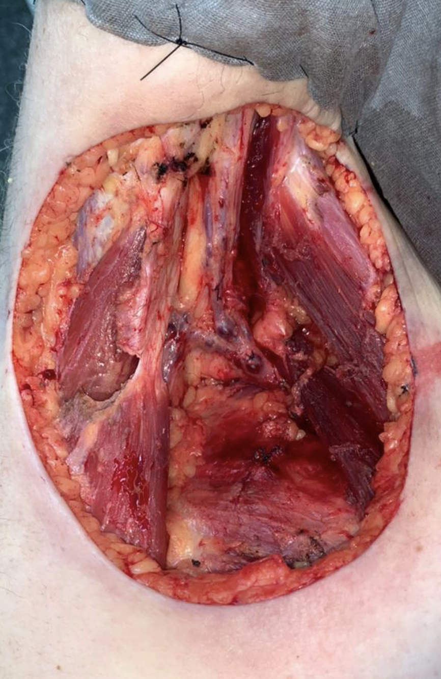
Conclusion: Despite the fact that the axillary area is not sun-exposed, squamous cell skin cancer manifested as an extensive lesion that required a complex surgical resection with flap repair. Such findings highlight the importance of a thorough physical exam and work-up to diagnose lesions in their early forms which require simple resection procedures and avoid late diagnoses resulting in complex procedures. Such an approach reduces the risk of various complications like wound infection or dehiscence, flap ischemia, or necrosis, among others.
References
Walter Belda Junior W, Di Chiacchio N, Criado P. Tratado De Dermatologia. 3rd ed. Rio de Janeiro: Atheneu Editora. 2018.
Ministério da Saúde do Brasil, Instituto Nacional de Câncer José Alencar Gomes da Silva (INCA). Estimativa 2020: incidência de câncer no Brasil. Rio de Janeiro: INCA. 2019.
Schmults CD, Blitzblau R, Aasi SZ, et al. NCCN Guidelines® Insights: Squamous Cell Skin Cancer, Version 1.2022. J Natl Compr Canc Netw. 2021;19(12):1382-1394. PMID: 34902824. https://doi.org/10.6004/jnccn.2021.0059
Stratigos AJ, Garbe C, Dessinioti C, et al. European interdisciplinary guideline on invasive squamous cell carcinoma of the skin: Part 1. epidemiology, diagnostics and prevention. Eur J Cancer. 2020;128:60-82. PMID: 32113941. https://doi.org/10.1016/j.ejca.2020.01.007
Schmults CD, Karia PS, Carter JB, Han J, Qureshi AA. Factors predictive of recurrence and death from cutaneous squamous cell carcinoma: a 10-year, single-institution cohort study. JAMA Dermatol. 2013;149(5):541-547. PMID: 23677079. https://doi.org/10.1001/jamadermatol.2013.2139
Kazantseva IA, Khlebnikova AN, Babaev VR, Fager G. Proliferativnaia aktivnost' bazal'nokletochnogo i metatipicheskogo raka kozhi [Proliferative activity of basal cell and metatypical cell carcinoma of the skin]. Arkh Patol. 1994;56(4):35-38. PMID: 7848103
Lin CT, Chen SG, Chen TM, Chang SC. Axillary basal cell carcinoma: case report and literature review. Acta Dermatovenerol Croat. 2011;19(2):107-109. PMID: 21703158
Huang CC, Boyce SM. Surgical margins of excision for basal cell carcinoma and squamous cell carcinoma. Semin Cutan Med Surg. 2004;23(3):167-173. PMID: 15584682. https://doi.org/10.1016/j.sder.2004.06.002
Tarallo M, Cigna E, Frati R, et al. Metatypical basal cell carcinoma: a clinical review. J Exp Clin Cancer Res. 2008;27(1):65. Published 2008 Nov 7. PMID: 18992138. https://doi.org/10.1186/1756-9966-27-65
Park SW, Heo EP, Choi JH, et al. Reconstruction of defects after excision of facial skin cancer using a venous free flap. Ann Plast Surg. 2011;67(6):608-611. PMID: 21372670. https://doi.org/10.1097/sap.0b013e318209a77f
Stratigos AJ, Garbe C, Dessinioti C, et al. European interdisciplinary guideline on invasive squamous cell carcinoma of the skin: Part 2. Treatment. Eur J Cancer. 2020;128:83-102. PMID: 32113942. https://doi.org/10.1016/j.ejca.2020.01.008
Published
How to Cite
Issue
Section
Copyright (c) 2022 International Journal of Clinical Research

This work is licensed under a Creative Commons Attribution 4.0 International License.